 440 Nicholson Rd, Forrestdale WA 6112
 (08) 9397 1286
 reception@dentalvet.com.au
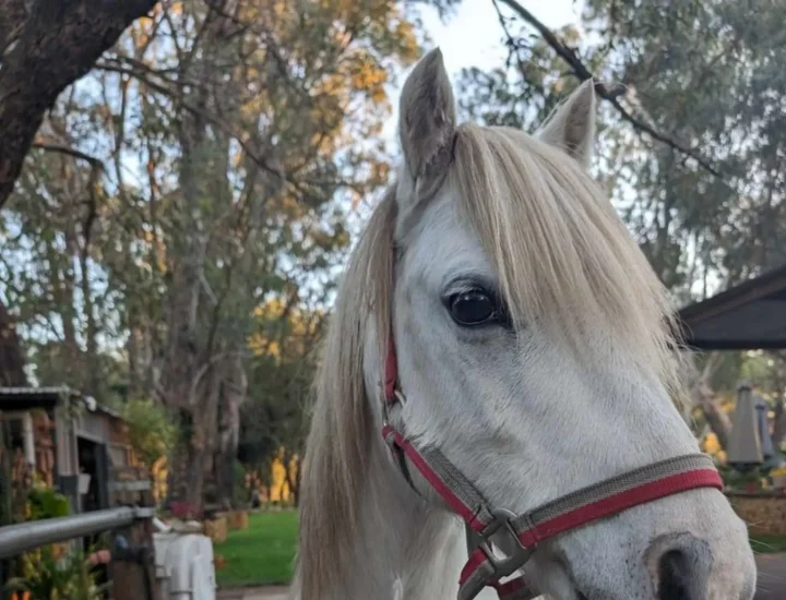
Caspa
Patient Information
Caspa the Welsh Pony's Journey with Mid-Sagittal Fractures
Patient History
Chewing Trouble Leads to Surprising Dental Diagnosis
Caspa, an eight-year-old Welsh Pony gelding, was initially booked for a routine dental examination. In the weeks prior, his owner had noticed he was struggling to eat hay and had developed firm swellings on both sides of his face.
Concerned, she arranged an appointment to investigate the cause of his discomfort.
Presenting Problem
Fractured Cheek Teeth and Severe Infundibular Decay
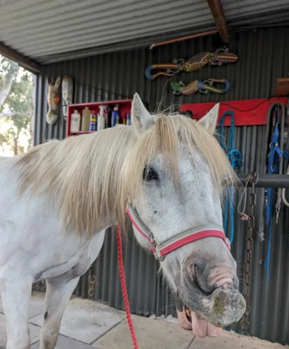
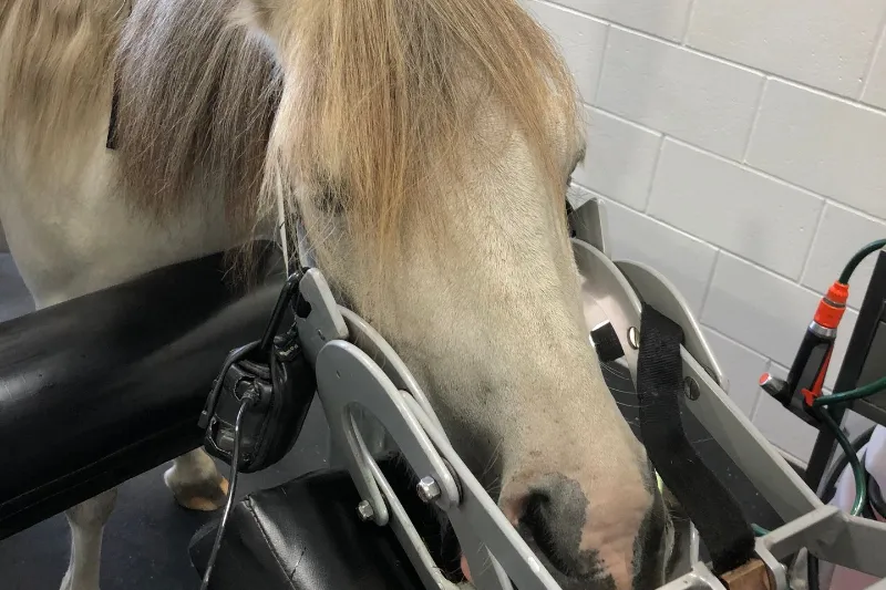
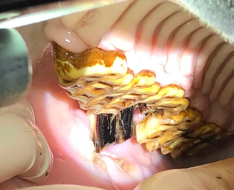
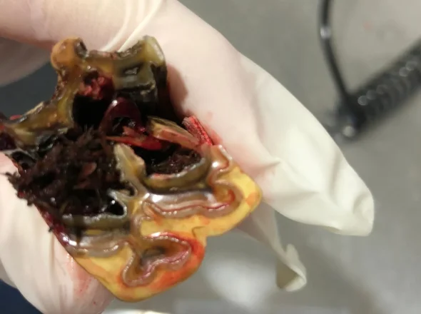
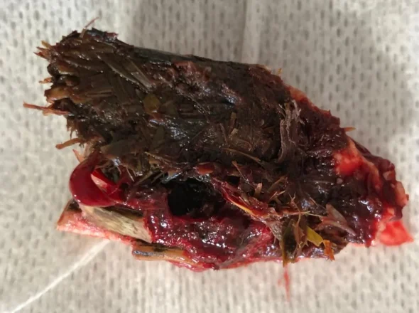
Under sedation and securely positioned in the stocks, Caspa underwent a detailed oral examination. The findings were striking. Both upper third cheek teeth (known as the 8s) had mid-sagittal fractures (teeth split vertically through the centre). The fractured fragments were protruding outward and ulcerating his cheeks, causing considerable pain.
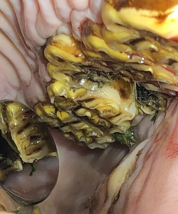
The fractures were secondary to a condition known as infundibular hypoplasia (underdevelopment of the central portion of the tooth), which leads to infundibular caries (decay in this central underdeveloped area). In this condition, the infundibula (the channels in the centre of the tooth) do not fill properly during development, leaving deep voids that trap feed and bacteria. Over time, the bacteria cause dental decay to progress, which weakens the tooth structure and, in severe cases like Caspa’s, can cause the tooth to split in half.
X-rays revealed that the teeth in the front (the 7s) were also affected by early infundibular decay, appearing less dense on imaging. Without intervention, these teeth were at risk of fracturing in the same way.

“Developmental infundibular defects can silently weaken the teeth for years before a catastrophic fracture occurs.”
Dr Kirsten Jackson
Diagnostics & Treatment
Extracting Fractured Teeth and Restoring Weak Teeth to Prevent Further Damage
At his first appointment, Caspa had three of the four fractured tooth fragments extracted. This provided immediate relief, as the sharp fragments had been ulcerating his cheeks. A follow-up appointment was scheduled a few days later to remove the remaining fragment safely, allowing for extended procedural time.
Once the patient was comfortable and healing well, attention turned to the teeth in the front, which were showing the same pattern of infundibular decay that had caused the earlier fractures. After discussing the risks of disease progression without treatment with his owner, it was decided to perform restorations (fillings) to seal the cavities and prevent further decay or fracture.
A few weeks later, Caspa was admitted to the Dental Vet clinic for the procedure. He was sedated and placed on constant-rate infusion to maintain stable sedation and analgesia throughout. IV fluids were administered to keep him hydrated and reduce the risk of post-operative complications.
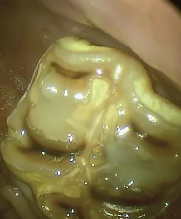
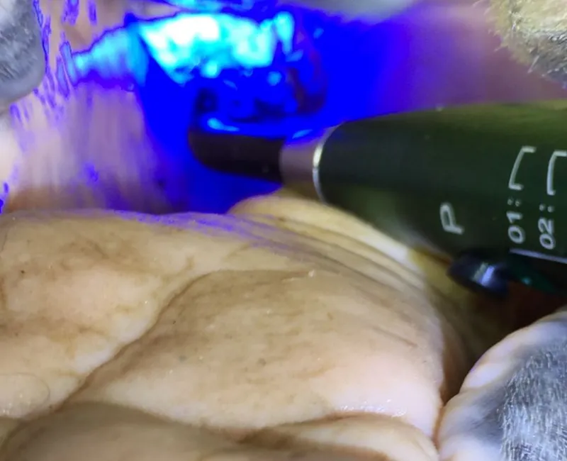
Each decayed infundibulum was thoroughly cleaned to remove packed feed and bacteria. Using a combination of dental drills, endodontic files, and cleaning medicaments, feed material was cleared from cavities as deep as 4cm. The infundibula were disinfected, etched, bonded, and then filled with a resin composite restorative material.
The restorative compound mimics the hardness of natural dentine, allowing it to wear naturally as the tooth erupts. This effectively completes the job the tooth’s own development had left unfinished. Once the restorations were set, the treated teeth were polished and equilibrated to ensure proper occlusion and comfort.
The owner was advised to monitor Caspa’s eating habits, avoid sugary feeds, and maintain routine dental checks to prevent recurrence.
“Restoring the infundibula stops feed and bacteria getting in, preventing the decay that leads to painful fractures.”
Dr Kirsten Jackson
Outcome
Lasting Comfort and Long-Term Tooth Preservation
Following the extraction of the fractured teeth, Caspa’s comfort improved immediately. He resumed eating normally, showing no further signs of pain or swelling.
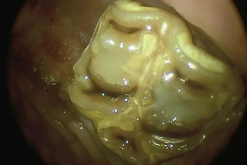
Years later, the results of his restorations remain excellent. The fillings are still intact, the decay has not progressed, and the previously weakened teeth have stayed strong and functional. Caspa continues to enjoy his hay and treats without discomfort. It’s the perfect outcome for a pony who once struggled to chew.
“Many years on, Caspa’s restorations remain in place and his teeth are healthy. He is proof that early intervention prevents long-term problems”
Dr Kirsten Jackson

Meet the Author
Dr Kirsten Jackson is the Owner and Director of Dental Vet and is passionate about her craft, treating every patient with the time, care and consideration she would give her own.
She is dedicated to improving the comfort and wellbeing of horses and other equine patients through advanced, preventative and compassionate dental care.
Key Takeaways
Infundibular hypoplasia can leave teeth structurally weak and prone to decay or fracture.
Restorative fillings can prevent future tooth loss and discomfort.
Developmental dental defects often remain hidden until advanced; early detection is vital.
Long-term success relies on regular monitoring and preventative dental care.

Advice for Owners
If your horse or pony drops feed, chews abnormally, or develops swelling around the cheeks, these may be signs of hidden dental problems. Routine dental examinations and early restorations can prevent painful fractures and preserve teeth for years to come.
FAQs
Do I need a referral?
No referral needed. In fact, we love to see your horse before any issues arise. Preventative dental care keeps your horse’s mouth healthy, so don’t put off going to the dentist. We do also accept referrals from veterinarians for advanced procedures, and will happily work with your vet to keep care seamless.
How do I book an appointment?
You can contact us directly to arrange an appointment on our online booking form or by emailing us at reception@dentalvet.com.au. Alternatively you can call (08) 9397 1286. We also work closely with referring veterinarians, sharing records and updates so your horse receives continuous care.
What’s included in a Dental Vet dental exam?
Every Dental Vet appointment is designed to give your horse a thorough, comfortable, and evidence-based dental assessment. Our experienced veterinarians take the time to thoroughly examine your horse, supported by our nursing team to ensure each visit runs smoothly.
Your horse’s appointment includes:
- A physical examination, including listening to the abdomen for signs of sand.
- A seven-point external head assessment to check for changes linked to dental disease.
- A comprehensive oral examination using a speculum, lights, and endoscopy to assess all teeth and oral tissues.
- Treatment of periodontal disease, if necessary (additional fee may apply for extensive treatment).
- Floating and corrective work, including reduction of sharp points and overgrowths, plus a performance float for bitted horses.
- Discussion of findings, next steps, as well as a dental chart emailed to you with home-care recommendations, so you understand each step.
- Optional bit fit assessment if you have bitting concerns.
Does Dental Vet accept pet insurance?
Yes, many equine insurance policies cover advanced veterinary dental treatment when performed by a registered vet. However, it’s always advisable to contact your pet insurer and check on your policy inclusions before the appointment.
What symptoms might indicate that my horse has a dental problem?
Horses are prey animals and instinctively mask their pain and discomfort, which means dental disease often goes unnoticed until it’s advanced. The signs can be extremely subtle, even the colour of the dentin overlying a 2mm pulp on the surface of the tooth can indicate whether a tooth is healthy or abscessed. By the time symptoms are noticeable, the problem may already be severe and require more extensive (and costly) treatment.
That’s why regular professional dental checks are so important, even if your horse seems completely normal.
If your horse shows any of the signs below, please contact us for a diagnostic examination as soon as possible:
- Dropping feed or slow eating
- Bit resistance or head tossing
- Weight loss or difficulty maintaining condition
- Nasal discharge or foul breath
- Quidding (spitting out half-chewed hay)
- Chewing on one side only
- Dunking feed or rinsing mouth in water trough
- Facial swelling or sensitivity to touch
What is your sedation policy?
Our patients are given a light, controlled sedation for their dental procedure. This keeps them calm and comfortable and ensures we can do a thorough examination, diagnostics and treatment. During our detailed examinations we assess over 140 pulp cavities, as well as infundibula and all dental and soft tissue structures with precision, while protecting both horse and handler. Each horse is individually assessed before sedation, and reversing agents and emergency medications are always on hand.

Whether you've got a new foal, referring a patient for review, or are worried about your horse's wellbeing, we're here to help!