 440 Nicholson Rd, Forrestdale WA 6112
 (08) 9397 1286
 reception@dentalvet.com.au
Facial swelling in horses: could it be a tooth root abscess?
10 March 2026 / Dr Kirsten Jackson
Tooth root abscesses or apical infections are a common, painful, often misunderstood and commonly undiagnosed dental issue in horses. Most cases develop quietly and show no outward signs. They are often only detected during a thorough oral examination with appropriate lighting, mirrors, or endoscopy, along with diagnostic imaging. External signs of a problem may include facial swelling, nasal discharge, or chewing on only one side.
Because the upper cheek teeth sit directly beneath the horse’s sinuses, infection around the tooth root can quickly affect surrounding structures and can result in a sinus infection with a smelly nasal discharge or a facial swelling in younger horses. This is often when owners first notice something is wrong.
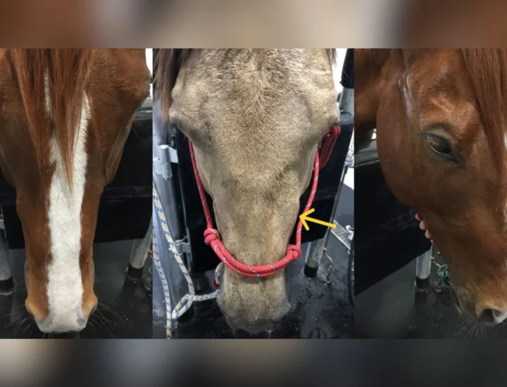
We treated a horse named Loki who presented with pronounced swelling along the side of his face. The infection had advanced from a diseased tooth into the surrounding bone, resulting in obvious external swelling.
However, not all cases are this obvious. In most horses, the disease develops quietly and is detected only during a thorough dental examination. Interestingly, facial swelling is actually less common than many people expect. It tends to occur more frequently in younger horses, where the teeth still fill much of the surrounding bone. In older horses, infection often drains into the oral cavity or sinuses, so the disease is not always visible externally.
What is a tooth root abscess?
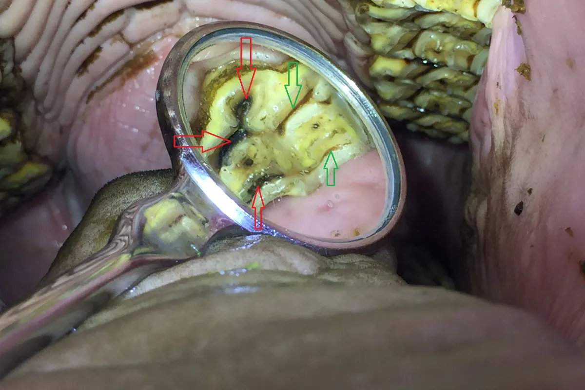
An infected tooth with a tooth root abscess. The red arrows point to open pulps, where feed and bacteria were entering the vital part of the tooth, indicating a likely dead tooth; the green arrows show what the normal protective layer over the pulps should look like. Also note the staining from the feed on the roof of the mouth is only on the opposite side, as she wasn't chewing on the affected side.
A tooth root abscess is an infection at the root of a tooth and involves the surrounding bone. Equine teeth have long reserve crowns extending well below the gum line. Understanding this helps explain how infections can spread and complicate quickly.
Infection from a diseased tooth can spread into the sinus or surrounding bone, leading to facial swelling or discharge from one nostril, or even an external draining tract where the infection drains through a hole in the skin.
Equine cheek teeth have complex anatomy, with multiple pulp canals that contain blood vessels and nerves in a healthy tooth. When infection enters these structures, it can overwhelm the immune system within the tooth and kill the vital part of the tooth. The infection can then quickly travel to the tooth root and bone surrounding the tooth, leading to apical infection or a ‘tooth root abscess’.
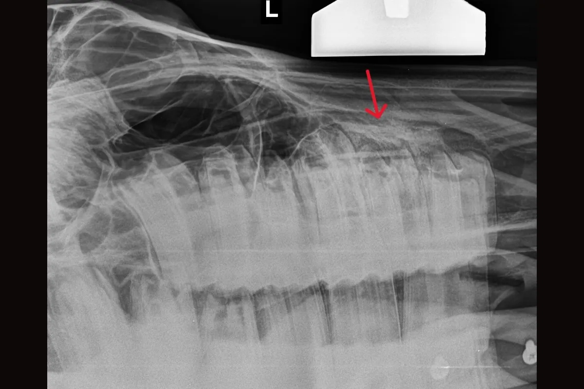
X-ray showing the affected tooth, with the arrow indicating the area of infection associated with the tooth root.
Why do tooth root abscesses develop?
While the process of a tooth dying usually occurs quite quickly after infection develops, it can take months or even years before any outward signs become visible.
Common causes include:
- Advanced dental disease.
Conditions such as infundibular caries or deep dental decay can extend into the pulp canals, allowing bacteria to enter the tooth. - Haematogenous spread.
Systemic infections can allow bacteria in the bloodstream to lodge in a tooth. If the infection overwhelms the immune system within the tooth, the tooth can die. - Periodontal disease.
Infection around the tooth can progress deeper into surrounding structures and eventually involve the pulp system, which can kill the tooth. - Tooth fractures.
Damage to the tooth may expose the vital tissue in the pulp canals, allowing bacteria to invade. - Development or structural abnormalities.
Some teeth have natural defects that make them vulnerable to disease.
Many of these problems develop slowly. Careful examination can often detect early disease before external signs appear. Identifying these subtle changes early can allow intervention before the infection progresses to the point of killing the tooth. Recognising these subtle changes is key to timely diagnosis and intervention, and, hopefully, to saving the tooth.
Early signs owners often miss
Many owners expect a tooth root abscess to present with obvious swelling or signs of pain. In reality, early signs are generally subtle, and there may be no external signs at all. Some diligent owners may notice slight changes in eating habits, mild oral discomfort, or minor facial asymmetry, making vigilance during regular health checks essential. In many horses, there may be no outward signs at all. The disease may only be detected during a detailed dental examination with appropriate imaging.
- Possible indicators include:
- Nasal discharge from one nostril.
- Mild swelling on the side of the face.
- Quidding or dropping partially chewed food.
- Slower eating, reluctance to chew hard feeds or careful placement of harder foods (such as a carrot) to avoid a painful area.
- Only chewing on one side.
- Changes in behaviour or performance under saddle.
However, it’s important to remember that most horses show no outward signs at all, or at least none that are noticed by the owner until the pain is relieved. Often, after the issue is resolved, many owners report a noticeable change in demeanour and attitude when the painful stimulus is removed (Pehkonen, J., Karma, L. and Raekallio, 2019). This reality underscores the value of diagnostic imaging and detailed oral examinations, even when a horse seems healthy.
One of our patients, Uno, appeared completely normal on the outside and showed no signs of discomfort or sinus disease.
During her routine dental examination, we noticed subtle changes on the tooth’s grinding surface, and, on further investigation with CT imaging, we identified a draining tract from an infected tooth. The infection was already advancing toward the sinus and was likely only days away from breaking through to cause a sinus infection.
Early detection allowed treatment before the condition became more complicated.
Often, the only sign is inside the mouth
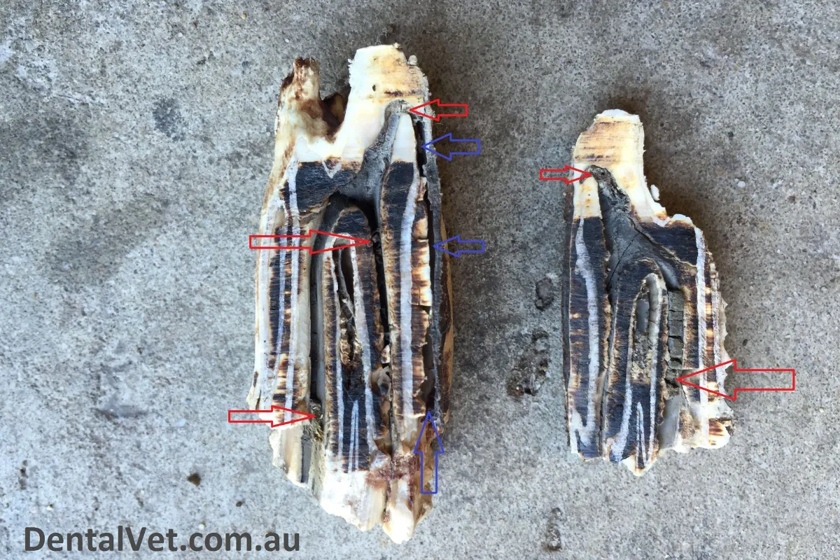
An extracted tooth with a tooth root abscess. The red arrows point to infected feed material within the pulp system, where blood vessels and nerves should be. The blue arrows indicate a draining tract through which infection was leaking back into her mouth.
In most cases, the only evidence of disease is subtle and only visible during a thorough oral examination, such as changes to the protective layer over the pulp cavities or faint radiographic changes. These changes can indicate that infection has entered the tooth’s internal structure and that the tooth has died.
In another case, the horse showed no obvious outward signs. There was no swelling or nasal discharge, and the owner hadn’t noticed any issues. However, during the oral examination, the protective layer over the pulp canals of one tooth appeared abnormal compared with those of neighbouring healthy teeth. Another subtle clue was visible on the roof of the horse’s mouth. Food staining was only present on the roof of the mouth on the opposite side, indicating that the horse was only chewing on the unaffected side.
When the tooth was extracted, pus could be seen bubbling from the gum beside the tooth. Once the tooth was sectioned, the pulp canals that should normally contain healthy blood vessels and nerves were instead packed with decaying feed material. A draining tract had also formed along the side of the tooth where infection had been leaking back into the mouth.
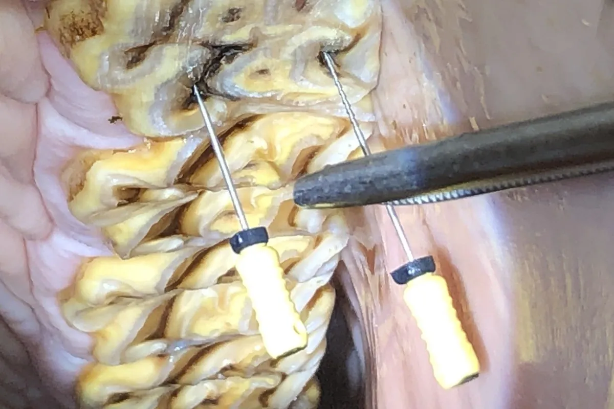
Tooth undergoing root canal treatment, with endodontic files used to clean and disinfect the infected pulp canals.
Treatment options
Treatment depends on the stage of the disease and the tooth’s structural condition. At Dental Vet, our goal is always to preserve the tooth whenever possible.
If the disease process is identified early, it may be possible to treat the infection before it progresses to the point of killing the tooth, allowing the tooth to be saved. This may involve treating severe areas of decay, including performing restorations (‘fillings’) to prevent the decay from entering the pulp system, treating acute pulp exposures in fractured teeth or addressing periodontal disease around the tooth before it progresses to irreversible pulp damage.
If the infection has entered the pulp system and the tooth is dead, we can, in many cases, perform endodontic (‘root canal’) therapy to save the functional tooth, provided the tooth is structurally sound. In these cases, we remove the infection within the tooth, fill the canals with a material that helps the bone heal, and seal them to prevent the infection from returning. In these cases, while the tooth is ‘dead’ (we can’t put new blood vessels and nerves in there!), it still functions and erupts as any normal tooth, preventing the secondary consequences of having a missing tooth after extraction. At Dental Vet, we are passionate about saving teeth and have spent many years researching and collaborating with human endodontists to develop new materials and techniques that improve patient outcomes. And we are hopeful that, once the work is published, the findings may also help horses around the world.
For this reason, extraction should generally be considered a last resort. Whenever the tooth structure allows, endodontic treatment can preserve normal dental function and avoid many of the long-term consequences associated with missing teeth.
In some cases, if the tooth structure is severely compromised, extraction may unfortunately be necessary to remove the source of infection and allow healing, but we certainly try to save teeth whenever possible.
If the infection has spread into the sinus cavity, additional treatment, such as sinus lavage, may be required to clear infected material and support healing. However, with early diagnosis and intervention, we can often treat before this happens and prevent more significant sequelae.
Follow-up examinations are also an important part of aftercare, as they allow healing to be monitored and help ensure the infection has resolved.
Long-term outlook
The long-term outlook for horses with tooth root abscesses is generally good when the condition is properly treated and the infection is resolved.
Most horses return to normal eating behaviour and performance once the affected tooth has been treated and the infection resolved.
If root canal therapy (RCT) has been performed, some ongoing monitoring is required to ensure that the pulps remain sealed and the infection resolves; however, if there is no other significant dental pathology, we usually only need to see these horses for their yearly routine dentals. This is in contrast to after a tooth is extracted, when horses generally recover well (barring complications); however, many sequelae require ongoing management for the rest of their lives. Horses’ teeth continue to erupt whether they have an opposing tooth to wear them down or not. So once a tooth is extracted, the opposite tooth will continue to overgrow and, if not maintained, will erupt through the opposite jaw. So more frequent (generally 6-monthly) dentals are usually required for the rest of their lives. Also, once a tooth is extracted, the teeth on either side migrate toward each other to close the gap. As the teeth move, this creates more gaps between the other teeth, allowing feed to pack into these gaps and secondary periodontal disease.
Having been performing RCT for 8 years now (and extractions for much longer!), we have had the privilege of watching these cases over many years, and it has been very interesting to observe the contrast between horses that have had a tooth extracted and those that have received RCT. After extraction, we are seeing the horses 6-monthly and always seem to be ‘putting out fires’, dealing with overgrown teeth, shifting teeth, periodontal disease and other issues. This is in contrast to just maintaining these healthy, normal arcades in RCT-treated horses and seeing them only once a year in most cases, so the long-term benefits to the horse (and the owner financially, with less frequent dentals!) can be considerable. Not every horse is a candidate for RCT, and in many cases, we have no choice but to extract the tooth. However, if we can save a tooth, we certainly try. Every tooth matters.
Early diagnosis allows treatment before infection spreads or more serious complications develop. With thorough examinations and modern diagnostic techniques, many infections can now be identified and treated early, preventing years of suffering, costly and invasive surgeries and improving animal welfare.
Reference: Pehkonen J, Karma L, Raekallio M. Behavioral Signs Associated With Equine Periapical Infection in Cheek Teeth. J Equine Vet Sci 2019;77:144-150.
Found this helpful? Subscribe to our newsletter for the latest
research, insights and updates.

Meet the Author
Dr Kirsten Jackson is the Owner and Director of Dental Vet and is passionate about her craft, treating every patient with the time, care and consideration she would give her own.
She is dedicated to improving the comfort and wellbeing of horses and other equine patients through advanced, preventative and compassionate dental care.

Whether you've got a new foal, referring a patient for review, or are worried about your horse's wellbeing, we're here to help!