 440 Nicholson Rd, Forrestdale WA 6112
 (08) 9397 1286
 reception@dentalvet.com.au
Equine Dental Anatomy: What’s Normal And What Isn’t?
Guide to Equine Dental Anatomy
At first glance, healthy horse teeth can look surprisingly rough, uneven and stained. That is often completely normal.
Unlike human teeth, equine cheek teeth are not designed to form a smooth, white surface. Their raised ridges, darker centres and shallow basins create the rough grinding surface needed to break down coarse, fibrous feed.
Most owners can see the incisors at the front of the mouth. The harder-working cheek teeth sit much further back. Assessing them properly requires sedation, a full-mouth speculum, strong lighting and a dental mirror or oral endoscope.
Understanding normal equine dental anatomy helps explain why we examine every surface carefully. It also explains why floating should never mean making the teeth look smooth.
Horse Teeth Wear And Erupt At The Same Time
Horse incisors and cheek teeth are hypsodont, meaning they have very long crowns. In a young horse, much of the crown remains below the gumline as reserve crown.
The tooth gradually erupts into the mouth as chewing wears away the exposed surface. Research suggests that cheek teeth generally wear at a rate of around 2-3 millimetres per year.
This is often described as continuous growth. However, once the ameloblasts have finished laying down the enamel part of the tooth, they die off, meaning once the tooth is formed, no more enamel can be produced. So while the teeth continue to erupt, and dentin and cementum do continue to be produced to allow for this continued eruption, the teeth actually get shorter with age!
Understanding that a tooth has a finite lifespan matters because every reduction removes tissue that cannot be replaced. Some occlusal work is needed in almost every horse to address sharp points, overgrowths, malalignment or changes contributing to periodontal disease. However, it should be targeted and limited to what is clinically necessary.
Indiscriminately flattening the natural ridges does two kinds of harm. It removes the textured surface the horse needs to crush fibrous feed, much like trying to cut hay between two sheets of glass. It also uses up more of the tooth’s finite crown, potentially shortening its functional life and making effective chewing more difficult as the horse ages.
Three Tissues Create The Grinding Surface
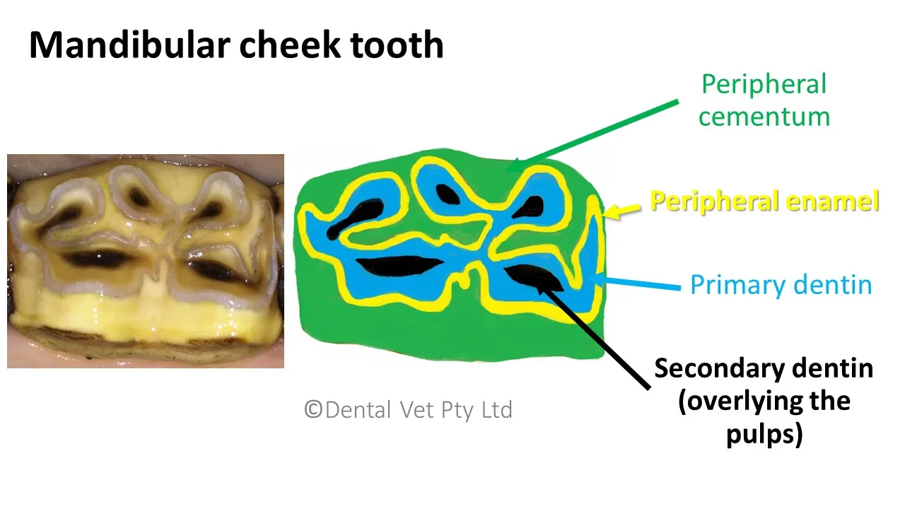
A healthy cheek tooth is closer to a serrated grinding plate than a polished table. Its working surface contains enamel, dentine and cementum, which wear at different rates.
Enamel is the hardest and most wear-resistant tissue. It runs through the tooth in complex folds. As the surrounding tissues wear more quickly, enamel remains slightly raised.
These ridges act as cutting edges during chewing. The lower areas between them help compress and break apart the feed. Together, they form the functional grinding surface. Dentine is softer and forms much of the tooth’s body. It supports the enamel and protects the living pulp system beneath the grinding surface.
Small brown areas on the surface commonly represent secondary dentine covering the pulp horns. These marks are normal when they remain firm, intact and appropriately coloured. A black, soft, open or irregular area is different. This may be the only detectable sign of a dead, infected, abscessed tooth.
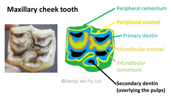
Cementum coats the outside of the clinical crown. It also fills the infundibula found within the upper cheek teeth and incisors.
Cementum is softer and less wear-resistant than enamel. It contributes to the self-sharpening surface, but it is also particularly vulnerable to demineralisation and dental caries.
What Are The Pulp Horns And Infundibula?
Inside each tooth is a living pulp system containing nerves, blood vessels and cells that produce protective dentine.
Equine cheek teeth contain between five and seven pulp horns beneath the grinding surface. Their connections and positions form a complex internal system that changes as the horse and tooth age.
Upper cheek teeth also contain two infundibula. These are deep, enamel-lined structures that should normally be completely filled with cementum.
On the grinding surface, healthy infundibula usually appear as light-yellow cement-filled areas surrounded by enamel. If the cementum is incomplete or missing, a cavity is left behind where feed can become trapped and ferment.
This fermentation gradually breaks down the remaining cementum, resulting in infundibular caries. Left untreated, the decay may progress deeper into the tooth, increasing the risk of fracture or, in advanced cases, infection involving the pulp.
Colour is one of several features we assess, but it is never considered in isolation. We also examine the depth and texture of the defect, the surrounding enamel, and its relationship to the pulp system to determine whether the change is within the range of normal anatomy or clinically significant disease.
What Should The Incisors Look Like?
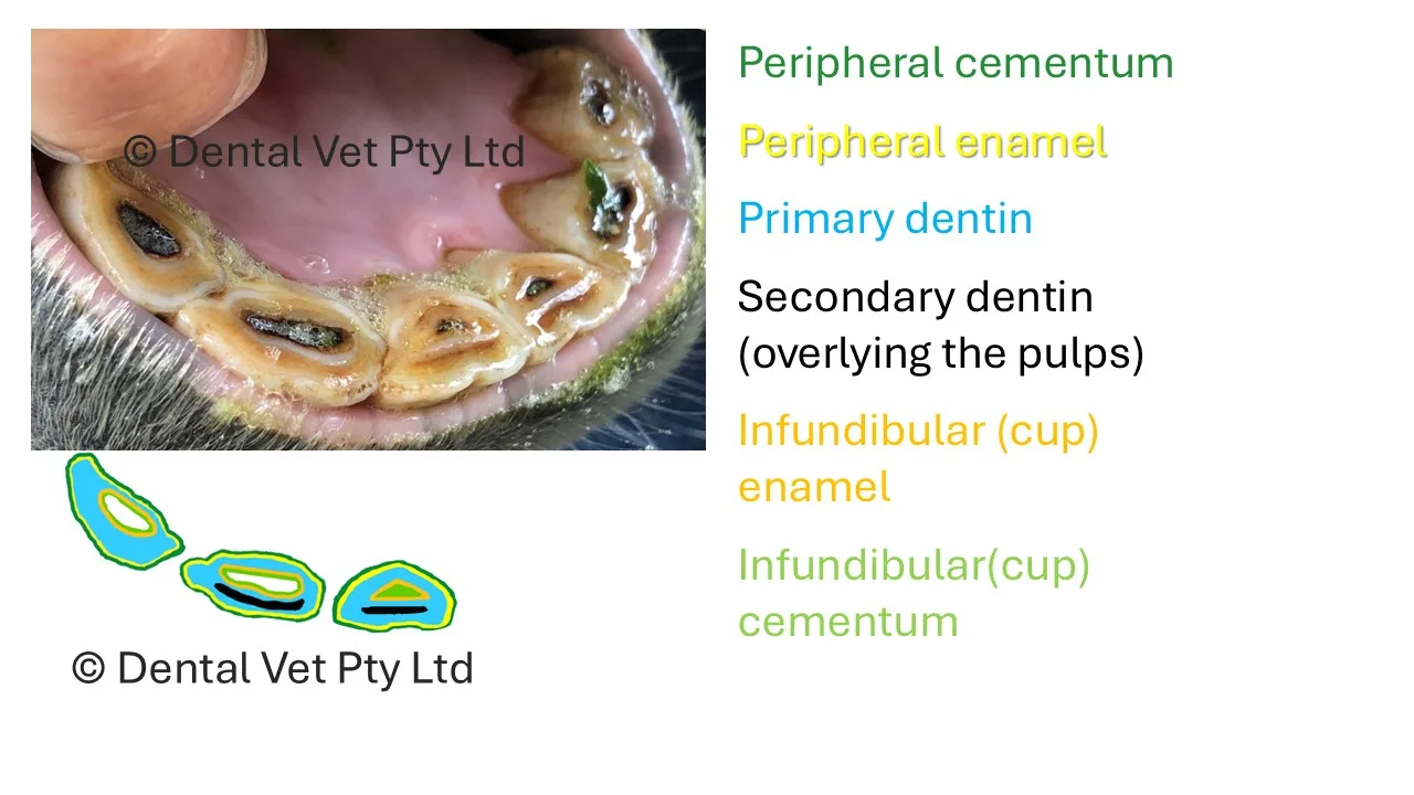
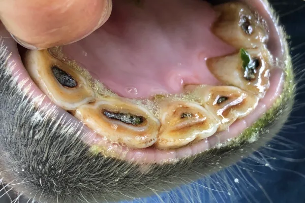
Incisors grasp and cut forage. Their visible surfaces change markedly as the teeth wear and age.
The cups within the incisors become shallower and eventually disappear. Dental stars, areas of secondary dentine that protect the pulp, become more visible.
The shape of the biting surface also changes. So does the angle at which the upper and lower incisors meet. Considerable variation can occur between individual horses.
These features are not automatically signs of disease. Fractures, marked mobility, draining tracts, swollen gums, severe asymmetry or painful resorptive lesions are different and require investigation.
A naturally dark dental cup should also not be confused with an exposed, infected or dead pulp. Both are large black holes in the teeth, but one is completely normal, and the other is likely a painful, infected tooth! This is why a thorough examination by a knowledgeable veterinarian is so essential.
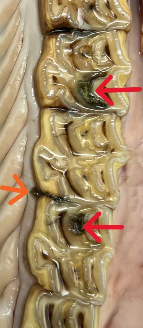
Arrows pointing to infundibular caries (red) and periodontal disease (orange)
What Does An Abnormal Tooth Look Like?
Some dental disease appears dramatic. We may find a large fracture, deep feed packing, a mobile or severely displaced tooth, severe decay or obvious gingival infection.
Other changes are tiny. A black pulp horn, a narrow fissure, missing cementum, or a subtle change in surface texture may be the only visible warning of some very painful dental disease.
Photographs are useful for education, but they cannot diagnose an individual horse. We must clean and examine the cheek teeth systematically under strong lighting.
We also assess the gums, the spaces between teeth, the cheeks, the tongue, and the remaining oral tissues. Where findings remain uncertain, radiographs or further diagnostic imaging may be required.
At our Forrestdale clinic, owners can view oral endoscope images while we explain what is normal and what requires monitoring or treatment. We also provide detailed dental charts, images and recommendations after the appointment.
Our aim is not to create a cosmetically perfect mouth. It is to preserve healthy anatomy, treat pathology and pain early and maintain comfortable, effective chewing throughout life.
That does not mean avoiding occlusal treatment. Quite the opposite. Correcting overgrowths, malalignment and other developing pathology at the right time often requires only small, targeted adjustments that preserve as much healthy tooth as possible. Left untreated, these changes often progress to more extensive problems requiring greater correction, ultimately shortening the functional lifespan of the affected teeth. By identifying and treating disease early, we can often preserve both the teeth and their function for longer, helping horses continue to chew comfortably well into old age.
Owners and referring veterinarians can contact our team by phone or email, or arrange an appointment online.
Found this helpful? Subscribe to our newsletter for the latest
research, insights and updates.

Equine Dentistry
Routine dental maintenance, including comprehensive examination of all dental and oral tissues as well as a full dental chart with recommendations.
Read More Read More
Lifetime Dental Care
Our vets will work with you and your horse on a comprehensive and tailored preventative dental care plan.
Read More Read More
Advanced Dental Procedures
Performing everything from endodontics, restorations, extractions and treating advanced periodontal disease.
Read More Read More
Equine Wellness Services
These include wellness consultations, vaccinations, microchipping, faecal egg counts (FEC), drenching, blood testing for metabolic disease, radiographs, euthanasia services and lameness work ups.
Read More Read More
In-Clinic Services
Take advantage of the state-of-the-art facilities our clinic has to offer. We are equipped with stables and yards with post, rail and mesh fencing and reticulated paddocks for your convenience. Clients can leave their horses for dental treatments, or stay and watch.
Read More Read More
Mobile Services
While we pride ourselves on our industry-leading clinic, we also bring premium care directly to your property when needed. Our mobile units are fully equipped to bring the same quality of care wherever you are.
Read More Read MoreFAQs
How can I tell if my horse’s teeth are healthy?
Healthy horse cheek teeth should have a rough, ridged grinding surface rather than a completely smooth one. The gums should not show significant inflammation, bleeding or deep feed packing. However, the back teeth require a sedated veterinary dental examination to be properly assessed.
Why do healthy horse teeth look rough?
Enamel wears more slowly than the surrounding dentine and cementum. This produces raised enamel ridges and lower basins that help shear and compress fibrous feed during chewing.
Are brown spots on horse teeth normal?
Small brown areas can be healthy secondary dentine protecting the pulp horns beneath the grinding surface. Black, soft, open or irregular areas may indicate pulp disease. We assess their colour, firmness, position and surrounding tooth structure before deciding whether they are normal.
Can a horse’s teeth be floated too much?
Yes. Excessive reduction may remove functional enamel ridges or damage the protective dentine over the pulp. Motorised instruments can also generate heat, so careful technique, cooling and conservative treatment are important.
Why do horse teeth change as they age?
Horse teeth gradually erupt as chewing wears away the exposed crown. Different levels of enamel, dentine and cementum become exposed over time, changing the tooth’s shape and grinding surface. These normal changes must be distinguished from decay, fractures and periodontal disease.
Can I check my horse’s back teeth myself?
You may notice obvious signs such as bad breath, quidding or facial swelling, but you cannot properly inspect the cheek teeth by lifting the lip. A complete examination requires sedation, a full-mouth speculum, strong lighting and dental examination equipment.

Google Rating
We love Kirsten from Dental Vet! She has been looking after Tinos teeth since he was 5 and he’s now 16. She is very kind and patient with Tino even when he can be a bit challenging and fidgety at times. Her advice and support has helped improve Tinos dental health over the years.
Thankyou Kirsten for a top job every time! We really appreciate your service
Riana Furheart
Google Rating
Have only just started using the services of these professional, kind Dental Vets. Our first impression was very prompt communication, leading to prompt arrival time, and treating both of our very different horses with exactly what they needed,to get the job done, with what stood out to me as genuine kindness to our two babies.The teamwork between Dr Tory and her colleague was great and they worked together seamlessly and even when we panicked a little with a bit of bleeding they both stayed super calm yet i knew they where keeping there eyes on him.
Bethany Heslop

Google Rating
Kirsten has been my horses dentist since she started back in 2010 and I wouldn’t use anyone else. She shows professional, knowledgeable, and genuine care to each and every horse. Her meticulous consults and work are not only beneficial to your horse but also to you as an owner. Kirsten spends the time to explain what she is doing, what she has found, what you as an owner should look for and what you can do. The learnings I have had over the years have given me a better understanding of my horses and how valuable qualified and experienced dentists like Kirsten are. There are not enough words to describe what an amazing person she is and the service her and her team provide. Our family highly recommend Dental Vet to anyone.
Fiona Reynolds
Google Rating
Kirsten is wonderful with my horses – including a 15yo rescue mare with a distrust of all people who had never had her teeth done in her life. She has been looking after all of my horses for 10 years now and I truly wouldn’t use anyone else. She is gentle, very professional and extremely passionate about dentistry and her research. And being a qualified vet often comes in handy! Would recommend without hesitation 🙂
heather6129
Google Rating
Dr Kirsten began assisting my horse’s dental situation with my vets while we were regional, and is now taking care of us now we are metro. Very informative, thorough and kind. My boy is in good hands and is getting the best care. Highly recommend.
Beth Giles
Google Rating
Dr Kirsten has looked after the dental health and general well-being of my horses for 11 years. She is absolutely meticulous and has the most caring manner with both horses and humans. We are incredibly fortunate to have such a knowledgeable equine dental professional in Western Australia.
Jade Salpietro
Google Rating
I highly recommend Dr Kirsten and her team. My warmblood gelding has had several difficult and problematic teeth issues that Dr Kirsten worked through systematically and with great care not only for my horse but for myself to understand all his treatment options and best course of action.
Dr Kirsten is highly intelligent, well read and passionate in her field and gives you 150% every appointment.
Sheree Jones
Google Rating
AMAZING 🤩 we are so lucky to have such a knowledgeable dental vet here in Perth. I wouldn’t go anywhere else for my horses dental check up and needs. Dr Kirsten gives a top quality service and is beautiful with the horses. Highly recommend!
Donna Mccoll
Google Rating
An outstanding equine dental experience! Dr Jackson and her team provide an exceptionally thorough and professional service, taking the time to build relationships with each individual client (both human and equine!). They even allow for overnight accommodation for your horse if you don’t have the time to wait during the procedure. Last time I accidentally left a windsucking collar behind and they posted it back to me, free of charge 🙂 Highly recommended in all aspects!
Sarah Murray
Google Rating
Dr Kirsten is lovely and kind. Her good character extends into her work, and together with her amazing knowledge in this industry, you will not go wrong using her equine dental services.
Jesslyn Seet
Google Rating
Always extremely happy with Dr Kirsten. She is very knowledgeable, Professional & kind. My horses teeth have never been better cared for!
L H
Google Rating
I have been attending Team Dental Vet since 2010. Or more acutely they have been coming to see me and my horses and ponies. With their portable crush, gentle approach, attention to detail and willing to adapt to the needs of the animals. I couldn’t have asked for a better team to look after my equine family.
Emma Andrews
Google Rating
Kirsten and her Dental Vet team are wonderful. I’ve trusted her with all of my horses over the past 15yrs as I know without a doubt my horses will get the treatment they need.
Ingrid Stanway
Google Rating
Dr Kirsten Jackson has been doing my mare’s teeth since 2015 & I wouldn’t go to any other dentist. Not only does she do a fabulous job every time but she is a wealth of knowledge & always happy to answer questions and explain what she is doing. Would highly recommend her services.
Jodie Baxter
Google Rating
Dr Kirsten, Dr Tori and the lovely nurses have been caring for my horses for 5 years now and even though we have moved further away we will keep coming back for their fabulous services and dedication to thorough dental care. Highly recommend Dental Vet!
Zakara Duval
Google Rating
I cannot rate Kirsten and her team at Dental Vet highly enough. The Dental Vet staff provide a professional, yet caring, atmosphere during each consultation. Kirsten takes the time to explain all aspects of dental care to her clients, and answers any questions you may have with an educated and friendly manner. The genuine care given to your horse, whether a performance horse or an old sole, is second to none. I wouldn’t take my horses to any other equine dentist!
Eileen Srna
Google Rating
I’ve used Dental Vet for my herd for over a decade. I switched to Dr Jackson having participated my horses in the evolution of equine dentistry for years, and had a variety of disappointing experiences, some traumatic. Dr Jackson restored my trust and cared for my herd collaboratively so we could always do our best, particularly for the elders, and balance the cost. For her professionalism, generosity of heart, of knowledge and financially, I am eternally appreciative. Our family trusts her with our beloved horse family, and subsequently we trust her staff, who also have never let us down.
Incidentally, we are blessed to have such a notable and internationally recognised Equine Dental expert in Western Australia. Thank you for your service! XJayne Lavender #TheHeartOfHorsemanship
Jayne Lavender
Google Rating
Best equine dentist in Australia. Thorough, kind, efficient, and affordable! Incredible restoration work, top notch care, safe, quiet, and clean facility. Cannot recommend Kirsten and the Dental Vet team enough!!!
Nym W
Google Rating
Would recommend Kirsten and all her staff to any one. Fabulous on site and comes to me off site with great service. They are all so kind and gentle with myself and my boy and no question is too silly or dismissed. If you want the best for your fur baby then contact Kirsten asap.
Lorraine Delamotte
Google Rating
Dr Jackson has been Willow’s dentist for 5 years. She has always brought a standard with her second to none. She is a true specialist in this exciting and emerging field of Equine Dentistry. There is so much more to horses’ teeth than rasping sharp points…and once you’ve booked Dr Jackson you will find out exactly why!
Heidi Niland-Rowe
Google Rating
Such a professional service and team. They are an asset to the Veterinary industry and have helped me with all of my horses. The knowledge and care is 2nd to none.
Caris Tavelli
Google Rating
Outstanding care for my horses dental health. As a vet myself I am very fussy about who attends to my horses needs. I trust the team at Dental Vet 110%
Belinda Beynon
Google Rating
Progressive vet practice, highly trained veterinarians providing clinical dentistry and dental surgery. These vets really care and go the extra mile for your animal and you.
Marieke Van Den Enden
Google Rating
Kirsten and her team are not only incredibly professional, knowledgeable and skilled, they are also lovely and kind.
Serena Monks
Google Rating
Dr Kirsten is very thorough and patient with horses and owners. Always informing, educating and very helpful.
Simone Tichelaar
Google Rating
Dr Kirsten, Tara and her team were so gentle and kind with my half blind boy Freckles. Thank you so much for your love and care xxx
Valerie Jean
Google Rating
Dr Tory and nurse Meg provided kind, gentle and extremely thorough dental care for my horses. Excellent work 🙂
Pia Hare
Google Rating
A dental vet Wizz!. Always up front with what needs to be done. Offers alternative treatment options according to budget. Always has the horses best interest central to her care.
Kerry “Kezbo87”
Google Rating
Lovely staff very professional and highly skilled vets. My horses dental health is in good hands.
Cindy Mathews
Google Rating
Dedicated, highly educated, exceptional care! Don’t hesitate getting your horses seen by Dr Kirsten!
Mikhalla Middleton
Google Rating
Kirsten provides the highest quality care for your equine partner. Gold standard care which is hard to find elsewhere.
Sarah M
Google Rating
Absolutely amazing. We are so lucky to having such a caring Dental vet. Love Noeline, Cim & Matt. Xx
Noeline Epis
Google Rating
Dr Kirsten is top of her game with horse dentistry she has been seeing my horses and donkeys for years. Thank you.
Ann Spendley
Google Rating
World class facilities and treatment standards. Would highly recommend to any caring horse owner who wants the best for their horses!
Oliver Liyou
Google Rating
Professional, extremely thorough and caring service by Kirsten and the team! Highly recommend!
Samantha Soltoggio
Google Rating
Super knowledgeable and experienced equine dentists – highly recommended.
Chanelle
Google Rating
Full dental service at a very reasonable price. Highly recommended.
Minty Choc
Google Rating
Expert dental care that I can always trust with my horses
Rena MacFarlane
Google Rating
Very knowledgeable and amazing professional service
Nikita Stibbard
Google Rating
Great experience, great veterinarian
Heather Burkhardt
Google Rating
Wonderful service 10/10
Jasslyn Wicks
Google Rating
Brilliant and knowledgeable.
Rachael Mason

Whether you've got a new foal, referring a patient for review, or are worried about your horse's wellbeing, we're here to help!