 440 Nicholson Rd, Forrestdale WA 6112
 (08) 9397 1286
 reception@dentalvet.com.au
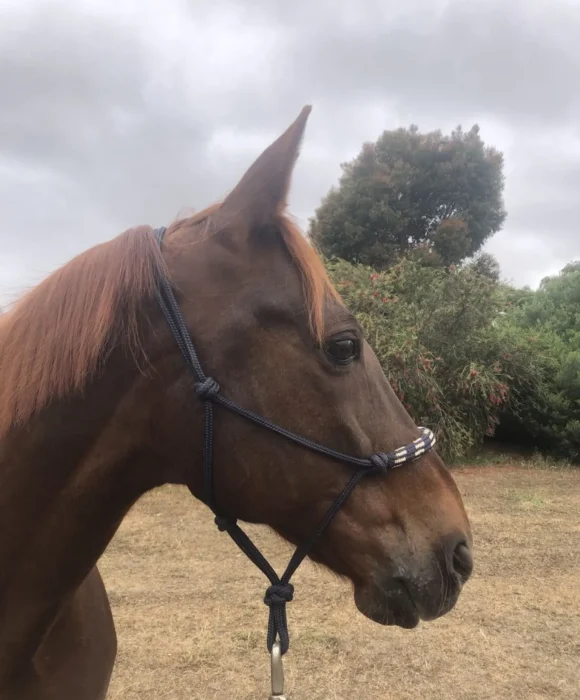
Jimmy
Patient Information
Jimmy the Standardbred gets Restorative Treatment for his Equine Dental Decay
Patient History
Routine Dental Exam Reveals Hidden Decay
Jimmy, a 13-year-old standardbred gelding, was booked in for his regular dental checkup. His owner had noticed some chewed-up balls of hay around the feed bin, known as quidding, and suspected something wasn’t quite right. While Jimmy was finishing his food and showed no major behavioural changes, the signs pointed to possible dental discomfort that warranted a closer look.
Presenting Problem
Extensive Caries and Significant Periodontal Disease Detected
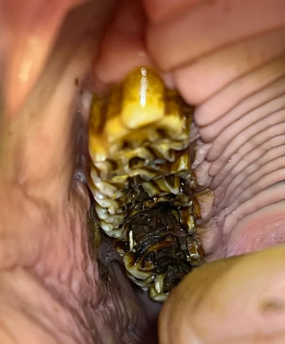
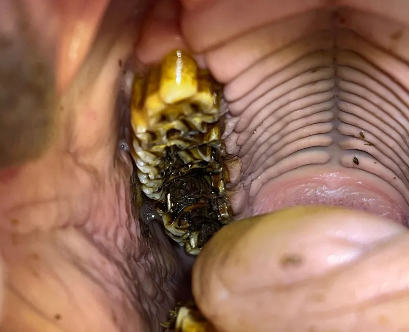
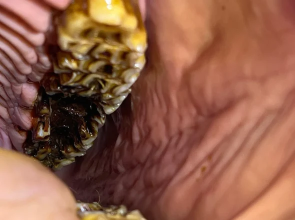
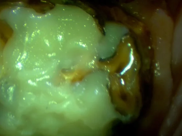
Jimmy was sedated and placed comfortably in the portable stocks for a full oral examination. The findings were significant. He had severe peripheral caries (decay on the sides of the teeth) and occlusal caries (decay affecting the grinding surface of the teeth) that had eaten deep into the protective layer over the pulp cavities (the inner chamber containing nerves and blood vessels).
The last three teeth in each arcade were the most severely affected, with much of the clinical crown (the visible part of the tooth above the gum line) eaten away to the level of the gum. Only small islands of enamel remained around the edges, forming perfect “caves” that trapped feed. This trapped material provided a constant food source for bacteria, accelerating decay and infection.
The peripheral caries had also eaten into the sides of the lower teeth, creating gaps where feed became wedged, causing secondary periodontal disease (infection between the teeth and under the gums). Several pockets of infection up to 10mm deep below the gumline were found.
Radiographs were taken to check for any tooth root abscesses that would indicate the decay had reached the pulp system. Fortunately, all roots appeared healthy, and treatment could proceed without extractions.

“Early detection is everything. Once decay breaches the pulp system, saving the tooth becomes far more complex”
Dr Kirsten Jackson
Diagnostics & Treatment
Comprehensive Restorative Dentistry to Halt Decay Progression
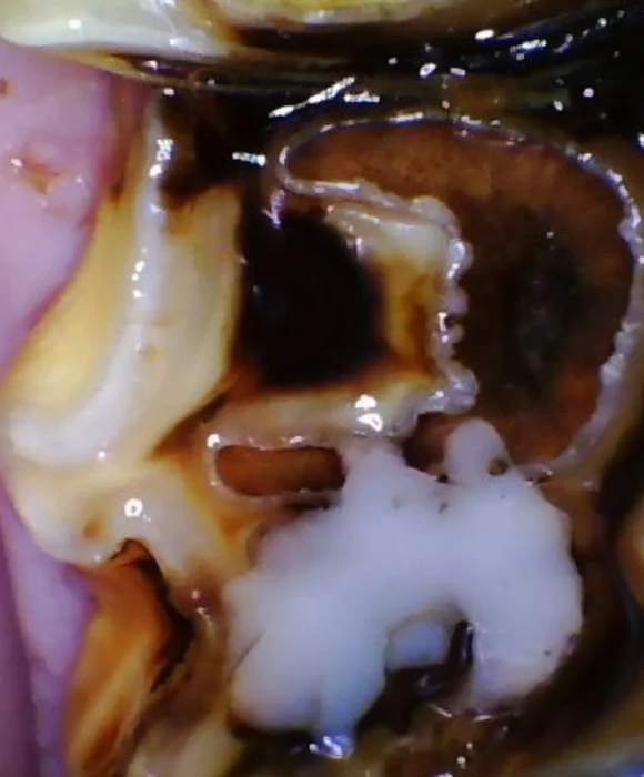
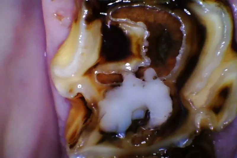
At the initial on-property appointment, treatment began by addressing the periodontal disease. All impacted feed material was removed, and the periodontal pockets were flushed with chlorhexidine antiseptic before antibiotics were packed into the infected spaces. Jimmy’s mouth was then equilibrated (balanced for even chewing pressure), and sharp enamel points were removed.
A fluoride varnish was applied to the areas of decay to help remineralise the dental materials and slow further damage until a follow-up appointment could be scheduled. Given the extent of decay, occlusal restorations (fillings) were recommended to prevent the infection from spreading into the pulp cavities.
At his follow-up appointment at the Dental Vet clinic in Forrestdale, Jimmy was sedated and placed under a constant-rate infusion for steady sedation and pain relief. He also received IV fluids to stay hydrated and minimise the risk of post-operative complications.
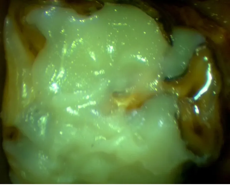
Eight teeth required restorations. The severely decayed material was carefully removed using precision dental drills until only the stronger, healthy dental material remained. The tooth surfaces were then shaped to create small ledges for retention (macromechanical retention), acid-etched, rinsed, and bonded. Finally, bulk-fill resin composite, a durable human dental material, was applied to restore the tooth’s shape and function.
These restorations served several purposes:
- Protected the pulp cavities to prevent further decay and pulp exposure, which would kill the tooth.
- Stopped food from becoming trapped, halting bacterial growth.
- Recreated a functional grinding surface for improved chewing.
- Provided immediate relief from dental sensitivity and pain.
The owner was advised to reduce sugars and cereal hays in Jimmy’s diet to prevent recurrence, as these feed types contribute to bacterial overgrowth and caries formation.
“Restorations don't just repair damage and prevent progression of the condition, they protect the living part of the tooth and provide instant relief from pain.”
Dr Kirsten Jackson
Outcome
A Happier and More Relaxed Horse
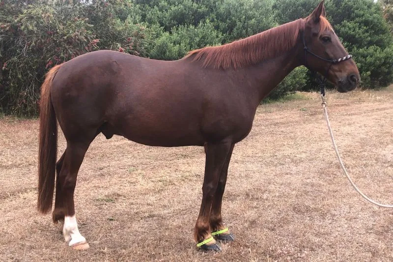
Two years after treatment, Jimmy’s progress was remarkable. The restorations had worn naturally with his teeth, and most of the damaged dental material had been replaced by a healthy new tooth as it erupted. Only small areas of filling remained.
His periodontal disease had resolved completely, and he was eating comfortably with no further signs of quidding or discomfort. His owners described him as a much happier horse, relaxed, bright, and back to enjoying his feed without hesitation.
“With timely treatment, even severe decay can be reversed as the new healthy tooth under the gum erupts. Jimmy’s recovery is a perfect example of why intervention matters.”
Dr Kirsten Jackson

Meet the Author
Dr Kirsten Jackson is the Owner and Director of Dental Vet and is passionate about her craft, treating every patient with the time, care and consideration she would give her own.
She is dedicated to improving the comfort and wellbeing of horses and other equine patients through advanced, preventative and compassionate dental care.
Key Takeaways
Caries can progress silently until extensive damage has already occurred.
Early intervention can save teeth that might otherwise be lost.
Restorations protect the tooth’s living structures and restore function.
Preventative care and dietary management reduce the risk of recurrence.

Advice for Owners
If your horse drops feed, forms quids, or leaves chewed hay behind, it could indicate dental discomfort. Regular dental checks are vital for early detection and prevention of painful conditions such as caries and periodontal disease.
FAQs
Do I need a referral?
No referral needed. In fact, we love to see your horse before any issues arise. Preventative dental care keeps your horse’s mouth healthy, so don’t put off going to the dentist. We do also accept referrals from veterinarians for advanced procedures, and will happily work with your vet to keep care seamless.
How do I book an appointment?
You can contact us directly to arrange an appointment on our online booking form or by emailing us at reception@dentalvet.com.au. Alternatively you can call (08) 9397 1286. We also work closely with referring veterinarians, sharing records and updates so your horse receives continuous care.
What’s included in a Dental Vet dental exam?
Every Dental Vet appointment is designed to give your horse a thorough, comfortable, and evidence-based dental assessment. Our experienced veterinarians take the time to thoroughly examine your horse, supported by our nursing team to ensure each visit runs smoothly.
Your horse’s appointment includes:
- A physical examination, including listening to the abdomen for signs of sand.
- A seven-point external head assessment to check for changes linked to dental disease.
- A comprehensive oral examination using a speculum, lights, and endoscopy to assess all teeth and oral tissues.
- Treatment of periodontal disease, if necessary (additional fee may apply for extensive treatment).
- Floating and corrective work, including reduction of sharp points and overgrowths, plus a performance float for bitted horses.
- Discussion of findings, next steps, as well as a dental chart emailed to you with home-care recommendations, so you understand each step.
- Optional bit fit assessment if you have bitting concerns.
Does Dental Vet accept pet insurance?
Yes, many equine insurance policies cover advanced veterinary dental treatment when performed by a registered vet. However, it’s always advisable to contact your pet insurer and check on your policy inclusions before the appointment.
What symptoms might indicate that my horse has a dental problem?
Horses are prey animals and instinctively mask their pain and discomfort, which means dental disease often goes unnoticed until it’s advanced. The signs can be extremely subtle, even the colour of the dentin overlying a 2mm pulp on the surface of the tooth can indicate whether a tooth is healthy or abscessed. By the time symptoms are noticeable, the problem may already be severe and require more extensive (and costly) treatment.
That’s why regular professional dental checks are so important, even if your horse seems completely normal.
If your horse shows any of the signs below, please contact us for a diagnostic examination as soon as possible:
- Dropping feed or slow eating
- Bit resistance or head tossing
- Weight loss or difficulty maintaining condition
- Nasal discharge or foul breath
- Quidding (spitting out half-chewed hay)
- Chewing on one side only
- Dunking feed or rinsing mouth in water trough
- Facial swelling or sensitivity to touch
What is your sedation policy?
Our patients are given a light, controlled sedation for their dental procedure. This keeps them calm and comfortable and ensures we can do a thorough examination, diagnostics and treatment. During our detailed examinations we assess over 140 pulp cavities, as well as infundibula and all dental and soft tissue structures with precision, while protecting both horse and handler. Each horse is individually assessed before sedation, and reversing agents and emergency medications are always on hand.

Whether you've got a new foal, referring a patient for review, or are worried about your horse's wellbeing, we're here to help!